

What is Image Enhanced Endoscopy (IEE)
Image Enhanced Endoscopy (IEE) holds various techniques that allow the endoscopist to better differentiate normal from abnormal tissues. This enables early detection of cancerous and precancerous areas to facilitate treatment.
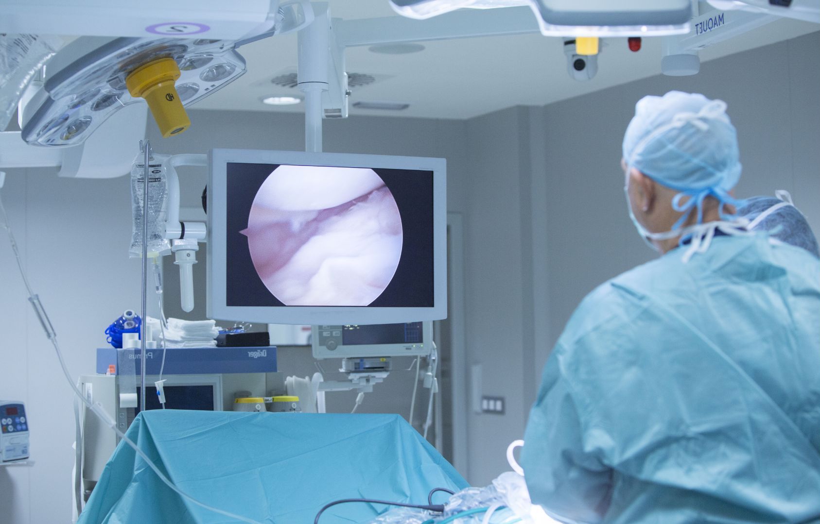
IEE highlights the microstructure and microvascular features of the mucosa or inner gut lining to enable accurate detection and delineation of cancerous and precancerous areas.
Broadly, there are two types of IEE: dye-based IEE and equipment-based IEE or also known as Digital Chromoendoscopy.
PROCEDURE
Dye based Chromoendoscopy
Dye-based IEE involves the application of various dyes to better identify abnormal tissues. These dyes are harmless to the human body and are sprayed onto the area of interest during endoscopy. The dye improves visualization of the microstructure and blood vessel pattern in the area of interest.
The main purpose is to clearly delineate the boundaries of cancerous and precancerous areas and to facilitate biopsies and endoscopic removal. The type of dye used depends on the location and type of cell that the endoscopist is looking for.
Chromoendoscopy has been proven in multiple studies to provide superior detection of precancerous or cancerous tissue compared to conventional endoscopy, particularly in individuals at high risk of cancer.
Digital Chromoendoscopy
Equipment-Based IEE is also known as Digital Chromoendoscopy. These are technologies where advanced imaging is incorporated into the endoscopy systems using either special light filters or computer algorithms to enhance surface features or micro-vascular characteristics. This eliminates the need for dyes, which is more time consuming and can only be used one at a time.
QUALITY Of The Procedure
IEE techniques allow detection of early cancers and pre-cancerous areas that routine endoscopy would fail to detect sometimes. IEE can be incorporated into routine endoscopy to provide a more thorough and detailed endoscopic examination. This facilitates cancer risk assessment and prevention for every patient undergoing endoscopy.
Accurate application of IEE requires the doctor to undergo a special course in IEE after training in general endoscopy. The doctor is also required to have a large series of cases comparing his ‘optical biopsy’ diagnosis against tissue biopsy diagnosis by the pathologist to ensure accuracy and consistency. The doctor should welcome questions regarding their training, credentials, and experience. The doctor should also be able to discuss the different types of IEE, and the various scenarios to apply specific IEE techniques
